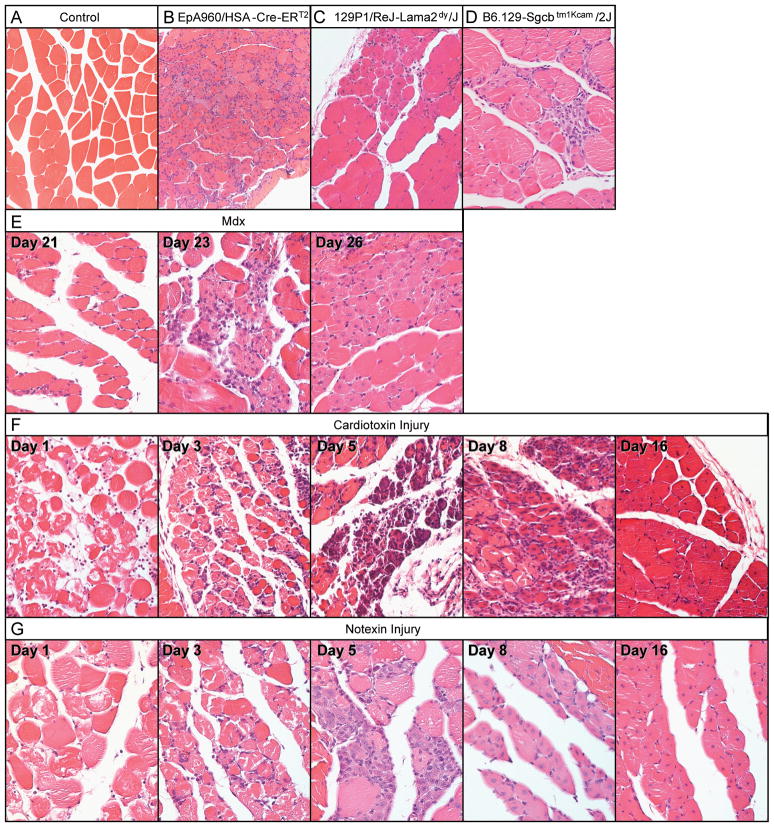
Figure 1. Histological analysis of six models of muscular dystrophy and muscle injury.
Hematoxylin and eosin stained cross-sections from paraffin embedded gastrocnemius muscle at 20X magnification. (A) Wild-type control, (B) EpA960/HSA-Cre-ERT2 (+ tam), (C) 129P1/ReJ-Lama2dy/J, (D) B6.129-Sgcbtm1Kcam/2J, (E) mdx at postnatal days 21, 23 and 26, (F) cardiotoxin and (G) notexin injured muscle at 1, 3, 5, 8 and 16 days post injection.

