Fig. 5.
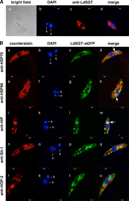
Colocalization studies by confocal microscopy. a Subcellular LdSGT localization in promastigotes by indirect fluorescence microscopy. Wild-type promastigotes were fixed and stained with DNA dye DAPI (b) and with anti-LdSGT (c) before visualizing by indirect immunofluorescence. A bright field image of the cell was obtained (a), the merged image (d) exclude the bright field figure. Bar = 5 µm. b Colocalization studies of LdSGT::eGFP with several chaperones and putative co-chaperones in fixed promastigotes. Wild-type promastigotes transfected with pTL-LdSGT::eGFP were fixed and stained with DAPI (b, f, k, o, s) and either anti-HSP70 (a), anti-HSP90 (e), anti-HIP (i), anti-Sti-1 (n), or anti-HOP-2 (r) and visualized by indirect immunofluorescence. C-terminal GFP-tagged LdSGT fusion protein was revealed by direct fluorescence (c, g, l, p, t). Overlays of direct GFP fluorescence, DAPI, and indirect immunofluorescence images are shown in bd, h, m, q, and u. The figure shows representative results of several independent experiments. Yellow color is indicative of colocalization. Arrows indicate clusters where the fluorescent patterns colocalize. Bar = 5 µm

