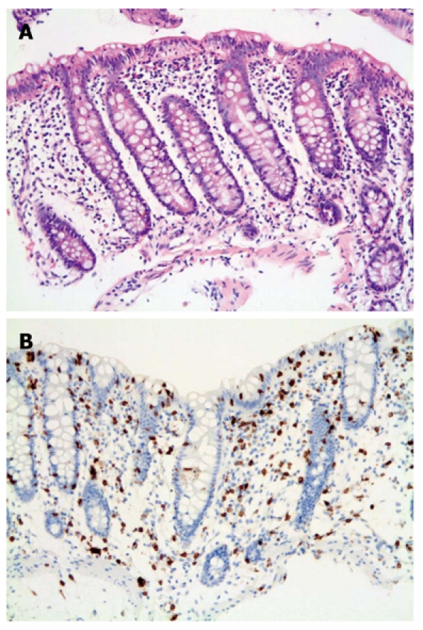
Figure 1.

Lymphocytic colitis. A: Classic form. Colonic biopsy showing typical findings of diffuse increase in intraepithelial lymphocytes, mild inflammation with surface epithelial damage (H and E stain × 200); B: CD3 immunohistochemistry highlighting lymphocytes (× 200).
