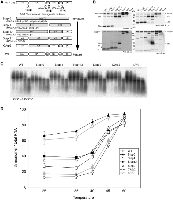
Figure 3.
The mutants to mimic sequential processing of HIV-1 Gag. (A) Schematic representations of the mutants. CA/p2 and NC/p1 mutants were added for reference. Shaded boxes represent fusion proteins which resulted from the mutations. (B) Detection of HIV-1 protein produced in pelleted virions by western blotting with various anti-Gag antibodies. Positions of Gag proteins and precursors are indicated. (C) Raw virion RNA profiles detected by northern blotting in a native agarose gel as in Figure 2. (D) Thermal dissociation kinetics of RNA dimers. The experiments were similarly performed as described in Figure 2. Results are the average of three to five independent experiments. Error bars represent SEM.

