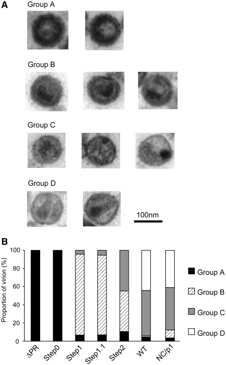
Figure 6.
Virion morphology of the mutants. (A) Classification of virion morphology judged by electron microscopy analysis. The criterion for judgment was summarized in Table 1. (B) The distribution chart of virion morphology in each mutant. A minimum of one hundred particles were surveyed and classified for each mutant.

