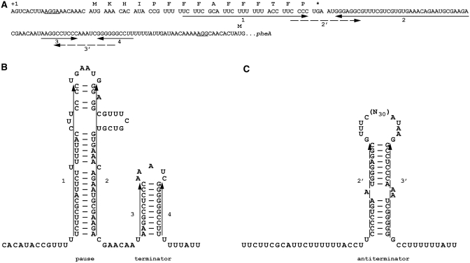
Figure 1.
Regulatory mRNA structures in pheA expression. (A) mRNA sequence of the phenylalanine operon from the transcription initiation site to the pheA initiation codon. The coding sequence of pheL is separated into codons with the encoded amino acids indicated above. Shine–Dalgarno sequences 5′ of the pheL and pheA initiation codons are underlined. Solid arrows indicate nucleotides involved in formation of the pause and terminator RNA structures. Broken arrows indicate sequences that form the anti-termination secondary structure. The run of ‘U’s at which transcription attenuates is in italics. (B) RNA secondary structures promoting attenuation of transcription. Transcribing RNA polymerase proceeds rapidly to a site just after RNA segment 2 where it pauses (44) due to formation of the 1:2 RNA secondary structure. With ample phenylalanine, ribosomes do not pause at the phenylalanine codons but proceed to the stop codon. Once RNA polymerase continues transcription, the 3:4 attenuator generally forms and transcription terminates at the 3′-run of ‘U’s. (C) RNA secondary structure favouring transcription and synthesis of pheA. When insufficient phenylalanine leads to aminoacyl-tRNAPhe becoming limiting and ribosome pausing at the run of 7 phenylalanine codons, RNA segment 2 is free to base pair with segment 3 which is transcribed once the polymerase is released from the pause. Formation of this antiterminator stem loop (2:3) permits transcription of the pheA coding sequence.

