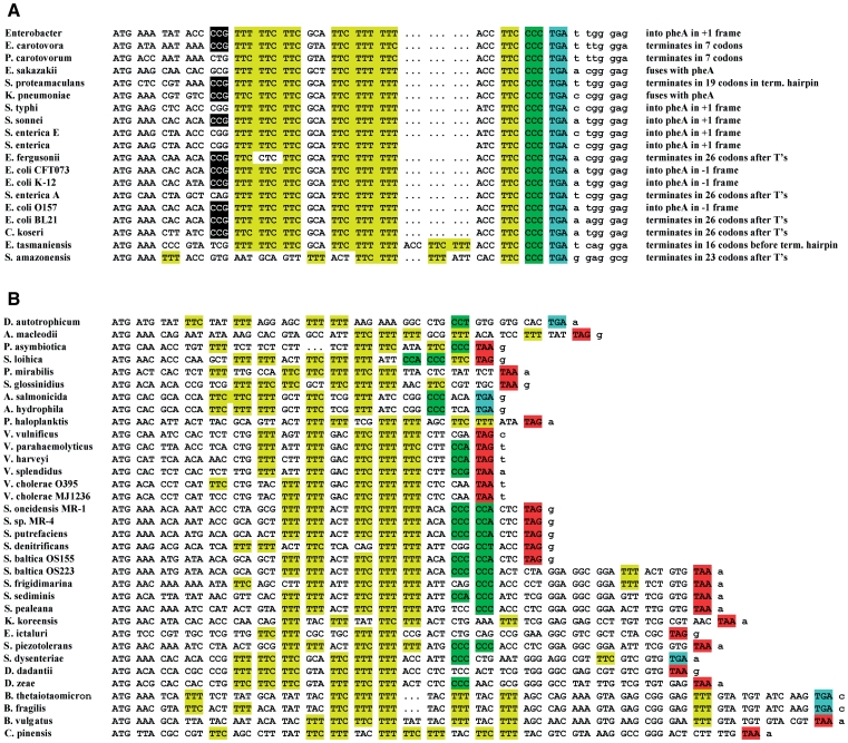
Figure 2.
The pheL sequences from different bacteria. When the nucleotide sequence is identical in several strains or species (e.g. E. coli K-12 and S. flexneri 2a) it is included only once in the alignment. Phenylalanine codons are highlighted in yellow; TGA stop codons—blue; TAA and TAG stop codons—in red; and Proline codons—in green. The sequence sources are in the supplementary material. (A) Alignment of the pheL sequences terminating with CCC_UGA. The position of the −10 Proline codon that has a 2-fold effect on frameshifting, is indicated by black shading. For each sequence, continuation of translation in the +1 frame after CCC_UGA is described in terms of where it terminates relative to the frameshift site, termination hairpin or attenuation site (run of ‘T’s) or whether it proceeds past the pheA initiation codon and in which frame. (B) Alignment of the pheL sequences that do not end with CCC_UGA.

