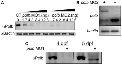
Figure 2.
Both polb morpholinos MO1 and MO2 can effectively block Polb translation which does not resume until 5 dpf. (A) Western blot showing loss of Polb protein after microinjection of different concentrations of MO1 (directed against the translation start site of polb) or MO2 (directed against the intron1/exon2 splice site). Protein extracts were prepared from 1 dpf (days post fertilization) embryos. (B) qRT–PCR shows that MO2, a splicing blocker targeting the intron 1/exon 2 junction, produced a polb splice variant that retains intron1 (upper band), besides the normal transcript (lower band). B-actin, shown in the lower panel, served as control. (C) Western blotting confirms that after knockdown of polb, Polb protein does not appear until 5 dpf. Total protein from Polb knockdown embryos and controls was extracted at 4 and 5 dpf, and examined by western blot analysis for the presence of Polb protein.

