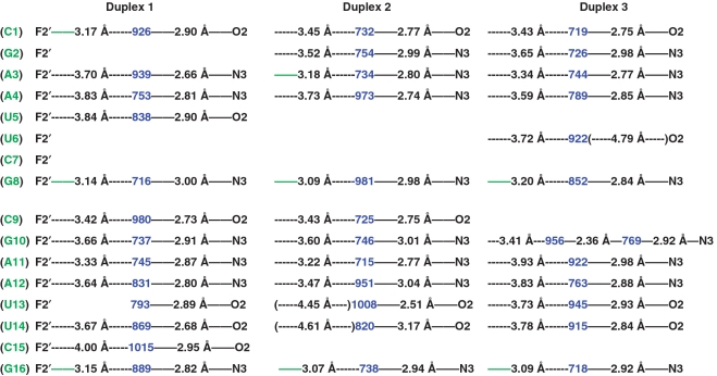
Figure 4.
Minor groove hydration pattern in the crystal structure of fA2U2 (three duplexes per asymmetric unit). 2′-F-RNA residues and water molecules (as numbered in the coordinate file) are highlighted in green and blue, respectively. Hydrogen bonds (≤3.3 Å distance between potential acceptor and donor atoms) are indicated with thin solid lines, with distances given in Å. Distances below 3.3 Å between fluorine atoms and water are highlighted in green. Please note the typically much longer distances (dashed lines in black) between 2′-fluorine atoms and water molecules in the minor groove of 2′-F-RNA compared with distances between nucleobase atoms and water or those between 2′-OH groups and water in RNA (Supplementary Figure S1).

