Figure 4.
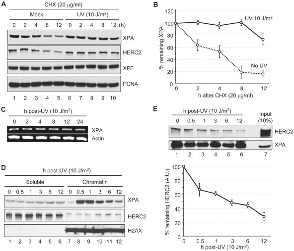
Effect of DNA damage on the interaction of XPA and HERC2. (A) Protein levels were analyzed by immunoblotting with the indicated antibodies. A549 cells were treated with UV or mock irradiated and then times were allowed as indicated for repair in the presence of cycloheximide (CHX). (B) Quantitative analysis of the XPA expression from the blot shown in panel (A). Averages and standard deviations were plotted from three independent experiments. (C) cDNA was prepared from cells allowed to repair UV damage for the indicated times and was used for PCR amplification of the genes indicated. (D) UV treated A549 cells were allowed to repair for the indicated times and cells were fractionated into soluble and chromatin fractions. Equal amounts of soluble and chromatin fractions were analyzed by immunoblotting with the indicated antibodies. H2AX was used as a chromatin loading control. (E) HERC2 interaction with XPA was analyzed by immunoprecipitation of XPA from UV treated A549 cells followed by immunoblotting of HERC2. Average values and standard deviations from three independent experiments are shown. HERC2 levels are shown as arbitrary units (AU) with 100% defined as HERC2 in XPA complexes from mock-irradiated cells.

