
Figure 5.
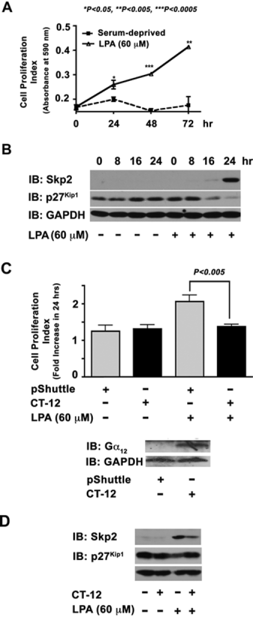
LPA upregulates Skp2 via Gα12. (A) Serum-deprived 1321N1 astrocytoma cells (5 × 10³ cells/well) were stimulated with 60 µM of LPA for 24 hours along with the unstimulated control group. The increase in cell numbers at 24, 48, and 72 hours was monitored using crystal violet–based dye binding assay as described in Materials and Methods. A fold increase at 24 hours was calculated to the absorbance at 0 hours. (B) Serum-deprived 1321N1 astrocytoma cells were stimulated with 60 µM of LPA for varying lengths of time (0, 8, 16, and 24 hours) along with unstimulated controls. Lysates (50 µg) from these cells were analyzed for the expression levels of Skp2 and p27Kip1 by immunoblot analysis using respective antibodies. The blot was reprobed with GAPDH antibodies to monitor equal protein loading. (C) Quiescent 1321N1 astrocytoma cells (5 × 10³ cells/well) infected with 150 MOI of CT-12 encoding adenovirus or empty pShuttle adenovirus (24 hours) were stimulated with LPA (60 µM) for 24 hours. Cell proliferation was measured using the XTT-based cell proliferation assay as described in Materials and Methods. Fold increase in cell proliferation was calculated by monitoring the increase in the absorbance of the formazan dye at 490 nm over a period of 24 hours. The expression of CT-12 was monitored by immunoblot analysis of lysates prepared from identical set-up experimental samples, using the Gα12 antibody that recognizes the CT-12 (lower panel). (D) Lysates (50 µg) from these cells were subjected to immunoblot analysis using Skp2, p27Kip1, or GAPDH. The results are from a representative set of experiments (n = 3).