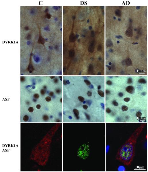
Figure 5.
Immunostaining of the frontal lobe cortex of control (C), Down syndrome/Alzheimer disease (DS), and Alzheimer disease (AD) subjects with mAb 7F3 illustrates comparable immunoreactivity for DYRK1A in the neurons of control and AD subjects, and greater immunoreactivity in DS/AD subject. There is no difference in the pattern of alternative splicing factor (ASF) immunoreactivity (mAb FS2/ASF) in the frontal cortex of control, DS/AD, and AD subjects. The third row shows a pyramidal neuron in the frontal cortex of a 67 y old DS subject with 3-color immunostaining. DYRK1A detected with mAb 7F3 is red, and ASF is green. Merged image with nuclei counterstained blue with TO-PRO-3I is shown in the right panel. Fine granular immunoreactivity for DYRK1A is partially colocalized with a fine granular ASF reaction product in the neuron nucleus.

