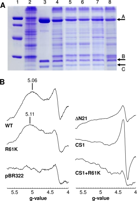
FIGURE 4.
A, SDS-polyacrylamide gel of DmsABC and its mutants. Lane 1, low molecular weight markers; lane 2, membranes from the negative control TOPP2/pBR322; lane 3, purified wild-type DmsABC; lane 4, TOPP2 membranes containing overexpressed wild-type DmsABC; lane 5, DmsAR61KBC; lane 6, DmsACS1BC; lane 7, DmsAΔN21BC; lane 8, DmsACS1+R61KBC. Arrow A marks DmsA (85.8 kDa), arrow B marks DmsB (22.7 kDa), and arrow C marks DmsC (30.8 kDa). 45 μg of total membrane protein was added per lane, except for lane 3, in which 30 μg of purified enzyme was used. B, effect of mutations of residues close to FS0 or the proximal pterin on the low field DmsABC EPR spectrum. EPR conditions were as described in the legend to Fig. 3, except that spectra are of membrane samples normalized to a protein concentration of 30 mg/ml.

