
FIGURE 2.
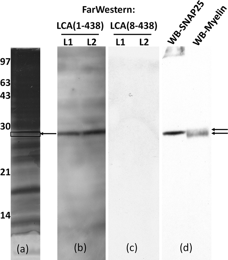
Far Western detection of LC/A bound to an ∼25-kDa protein in rat brain plasma membrane lysates. a, Triton X-100-extracted rat brain plasma membrane lysates were subjected to SDS-PAGE. Gel was stained with Coomassie Blue. The boxed area of the gel (arrow) was cut out and subjected to MS analysis as described in Table 1. b and c, the gel was transferred to PVDF membranes, blocked, and subjected to Far Western analysis, incubating with 0.1 μm 3xFLAG-LC/A(1–438) (b) or 3xFLAG-LC/A(8–438) (c) for 2 h. Bound LC/A was detected with α-FLAG-HRP antibody followed by SuperSignal analysis; an image of the x-ray film is shown (lanes L1 and L2 are identical lysate samples). The left blot was stripped, and the L1 and L2 lanes were divided and probed for SNAP-25 (α-SNAP-25 antibody) or myelin (α-myelin antibody), respectively. Bound antibody was detected with an HRP secondary antibody followed by SuperSignal analysis. d, an image of the x-ray film is shown. WB, Western blot.