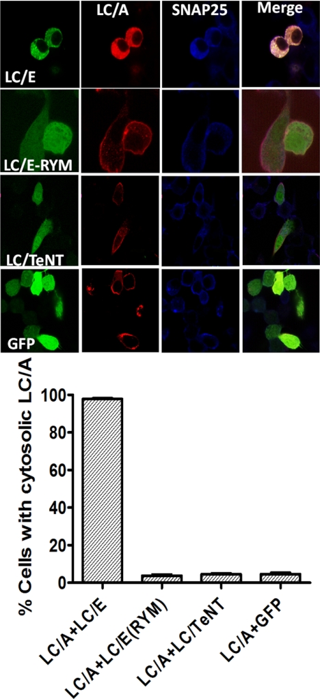
FIGURE 6.
Coexpression of LC/E disrupts LC/A plasma membrane localization. Upper panel, Neuro-2A cells were cotransfected with pLC/E, pLC/E(R347A,Y349F) (LC/E(RYM) pLC/Tetanus toxin (LC/TeNT), or pGFP derivative (green) and pLC/A (red). Cells washed, fixed, and probed for SNAP-25, using α-SNAP-25 IgG (blue). Cells were visualized by confocal microscopy, and images were merged. Lower panel, greater than 100 cells were scored for LC/A cytosolic phenotype under each transfection treatment. Results are the average of three independent experiments. Error bars indicate S.D.

