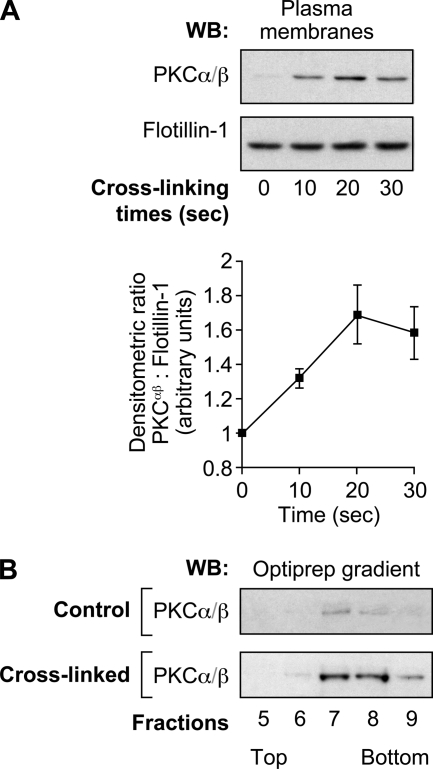
FIGURE 6.
PKCs translocate to the plasma membranes in DRM-Hs following FcγRIIa cross-linking. Neutrophils (40 × 106 cells/ml) were preincubated with 1 mm DFP for 10 min prior to FcγRIIa cross-linking at room temperature. A, plasma membranes were prepared as described under “Experimental Procedures” and analyzed by immunoblotting for the presence of PKCα/β. The same blot was reprobed for flotillin-1 (protein loading control). A densitometric quantification of the means of three independent experiments is shown in the line graph. B, plasma membranes of resting or FcγRIIa-cross-linked (30 s) neutrophils were prepared as described under “Experimental Procedures” and solubilized in 1% Nonidet P-40. These samples were subjected to ultracentrifugation on OptiPrep density gradients as described under “Experimental Procedures.” The 13 gradient fractions were collected, and the proteins were precipitated and analyzed by immunoblotting using the anti-PKCα/β antibody. These data are representative of three independent experiments. WB, Western blot. Error bars, S.E.

