FIGURE 2.
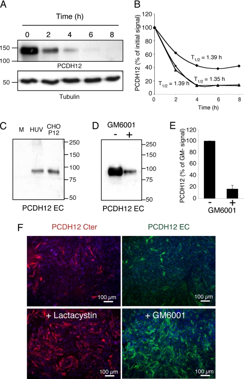
Instability of PCDH12 protein at the cell surface. CHO-PCDH12 were incubated in presence of cycloheximide (100 μg/ml). A, cells were harvested at indicated times, and protein extracts were analyzed by Western blotting using PCDH12 and tubulin antibodies. B, intensities of PCDH12 signals from three independent experiments were plotted, and protein half-lives (T½) were deduced. C, cell conditioned media of confluent CHO-PCDH12 (CHO-P12; 40 μl) or HUVECs (HUV; 40 μl after 40× concentration) were harvested after 24 h of incubation and analyzed by Western blot with anti-PCDH12 EC antibody. Unconditioned medium (M) was run as control. D, cell conditioned media of CHO-PCDH12, cultured for 24 h in presence (+) or absence (−) of 10 μg/ml of metalloprotease inhibitor GM6001, were analyzed as in C but with a longer exposure of autoradiographic film. E, quantitative analysis of three experiments performed as in D. Histogram shows the means ± S.D.; p < 0.001. F, images show PCDH12 immunofluorescence of CHO-PCDH12 cultured in regular medium or treated with either lactacystin 5 μm or GM6001 10 μg/ml, as indicated.

