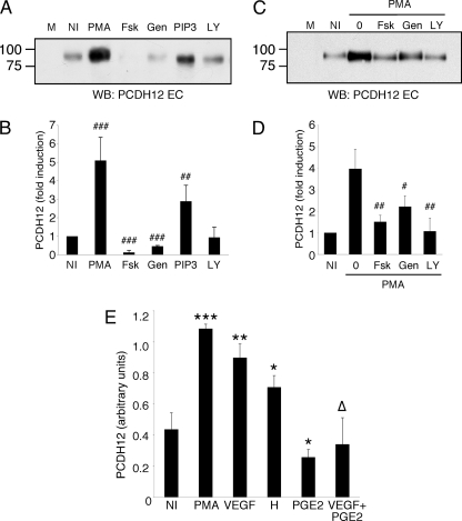
FIGURE 6.
PCDH12 shedding is regulated by various transduction pathways. A and B, CHO-PCDH12 were treated with Me2SO (NI), 0.1 μg/ml PMA, 10 μm Fsk, 50 μm genistein (Gen), 10 μm 1,2-dipalmitoylphosphatidylinositol 3,4,5-trisphosphate (PIP3), or 10 μm LY294002 (LY). The cell supernatants were harvested at 24 h and analyzed by Western blot, as illustrated in A. M stands for unconditioned cell medium. B, supernatants of three independent experiments were also analyzed by dot blot, and the signal intensities were measured. The data represent the means ± S.D. of signals normalized with signals of controls (NI). Statistical analysis compares controls versus treated cell signals. C and D, CHO-PCDH12 were treated with Me2SO (NI) or PMA alone (0) or PMA together with either forskolin, genistein, or LY294002. Cell supernatant were harvested and analyzed as above. Statistical analysis compares double treatments to PMA alone. ###, p < 0.001; ##, p < 0.01; #, p < 0.02. E, HUVECs were incubated with Me2SO (NI), 0.1 μg/ml PMA, 200 ng/ml VEGF, 1 mm histamine (H), 100 nm PGE2, or VEGF and PGE2. The supernatants were harvested at 24 h, and 100 μl were used for dot blot analysis. The data are the means ± S.D. of triplicates and are representative of three independent experiments. Significance with NI: ***, p < 0.001; **, p < 0.01; *, p < 0.05. Significance with VEGF alone: Δ, p < 0.01.

