FIGURE 3.
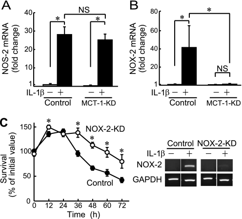
Involvement of NOX-2 in IL-1β-induced cell death. A and B, the expressions of mRNAs for Nos-2 (A) and Nox-2 (B) were quantitatively analyzed by real-time RT-PCR in Mct-1-silenced (MCT-1-KD) and control ATDC5 cells with or without treatment with IL-1β (10 ng/ml) for 48 h. Amplification signals from the target genes were normalized against that from GAPDH. The results are shown as values relative to that obtained from the control cells (far left column). C, Nox-2-silenced (NOX-2-KD) and control ATDC5 cells were plated at a density of 6.0 × 10⁴ cells/well and cultured for the indicated periods in the presence of IL-1β (10 ng/ml). Cell viability was evaluated using an MTT assay. ○, Nox-2-silenced cells. ●, control cells. Right, the expression of Nox-2 mRNA in control siRNA- and Nox-2 siRNA-introduced ATDC5 cells (NOX-2-KD) was assessed by RT-PCR after incubation for 48 h with IL-1β (10 ng/ml). A–C, results are expressed as the mean ± S.D. (error bars) of four independent experiments. *, p < 0.05. NS, not significant.

