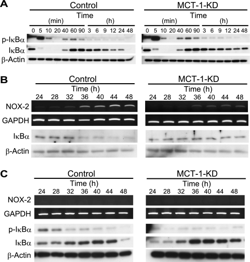
FIGURE 4.
Involvement of MCT-1 in IL-1β-induced IκBα degradation and Nox-2 expression. A, phosphorylation and degradation of IκBα after treatment with IL-1β (10 ng/ml) in control (left) and Mct-1-silenced (MCT-1-KD) ATDC5 cells (right) at various time points were examined by Western blotting. IκBα disappeared after 48 h in the control cells, whereas it remained in the Mct-1-silenced cells. B, a more precise study of the time courses of Nox-2 expression and IκBα degradation was performed in the period from 24 to 48 h after the addition of IL-1β in the control (left) and Mct-1-silenced (MCT-1-KD) ATDC5 cells (right). C, inhibition of IκBα degradation and Nox-2 expression by a proteasome inhibitor, MG-132. Control (left) and Mct-1-silenced (MCT-1-KD) ATDC5 cells (right) were treated for the indicated periods with IL-1β (10 ng/ml). MG-132 (5 μm) was added to the culture 4 h before each isolation of total RNA and protein fractions from cells at the indicated time points. The expression of Nox-2 mRNA was examined by RT-PCR, whereas those of phospho-IκBα (p-IκBα) and IκBα were assessed by Western blotting.

