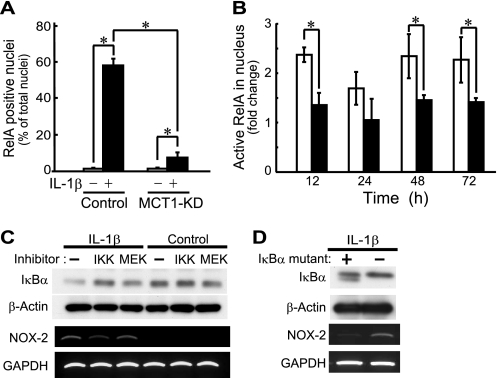
FIGURE 5.
Involvement of MCT-1 in IL-1β-induced activation of p65/RelA. A, the subcellular distribution of p65/RelA was examined by specific immunostaining at 48 h after treatment with IL-1β. The percentage of cells with p65/RelA localized in the nucleus is expressed as the mean ± S.D. (error bars) of four independent experiments. *, p < 0.05. B, control siRNA-introduced (unfilled columns) and Mct-1 siRNA-introduced (filled columns) cells were incubated with IL-1β (10 ng/ml) for the indicated time periods. Active p65/RelA in nuclear extracts was determined by ELISA-based quantification of p65/RelA bound to κB oligonucleotide. See “Results” for details. C, ATDC5 cells were cultured for 48 h in the presence or absence of IL-1β (10 ng/ml). At 24 h after the addition of IL-1β, an IKK inhibitor, BAY 11-7082 (2.5 μm) (IKK), an MEK inhibitor, PD98059 (50 μm) (MEK), or the vehicle alone (−) was added to the culture. Top panels, protein levels of IκBα and β-actin were assessed by Western blotting. Bottom panels, the expressions of mRNAs for Nox-2 and GAPDH were assessed by RT-PCR. D, cells were infected with retroviruses harboring an IκBα mutant, the NF-κB super repressor, or a control virus for 24 h. After selection of the infected cells by 24 h of incubation with puromycin, cells were further incubated for 48 h with IL-1β (10 ng/ml). Expressions of IκBα and β-actin (top) and those of Nox-2 and GAPDH (bottom) were assessed by Western blotting and RT-PCR, respectively.

