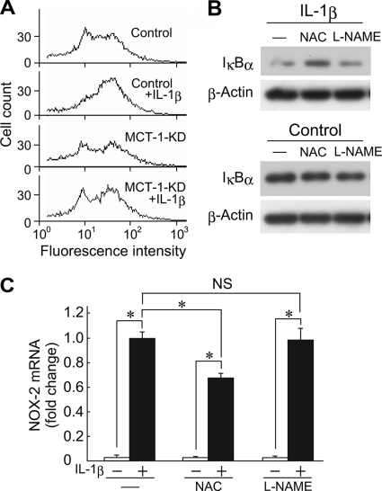
FIGURE 6.
Involvement of ROS in late phase activation of NF-κB after IL-1β treatment. A, control and Mct-1-silenced (MCT-1-KD) ATDC5 cells were cultured for 15 h in the presence or absence of IL-1β (10 ng/ml). The amount of ROS was measured using flow cytometry. B, ATDC5 cells were cultured for 48 h in the presence or absence of IL-1β (10 ng/ml). At 24 h after the addition of IL-1β, NAC (5 mm) or l-NAME (5 mm) was added to the culture. The protein levels of IκBα and β-actin were assessed by Western blotting. C, ATDC5 cells were cultured for 48 h in the presence or absence of IL-1β (10 ng/ml). At 24 h after the addition of IL-1β, NAC (5 mm) or l-NAME (5 mm) was added to the culture. The expressions of mRNAs for Nox-2 and GAPDH after 48 h of incubation with IL-1β were assessed by real-time RT-PCR. Amplification signals from the Nox-2 gene were normalized to that from the GAPDH gene. The results are shown as values relative to those obtained from cells exposed to IL-1β in the absence of NAC or l-NAME. Results are expressed as the mean ± S.D. (error bars) of four independent experiments. *, p < 0.05. NS, difference is not significant.

