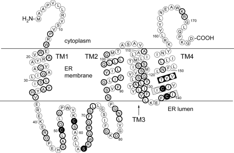
FIGURE 1.
A topological model for human VKORC1L1 based on sequence alignment (see supplemental Fig. S2) to a prokaryotic VKOR homolog protein structure determined to 3.6 Å resolution. Circles, represent amino acid residues; bold circles, indicate sequence identity shared by VKORC1L1 and paralog VKORC1; TM1–TM4, first through fourth transmembrane α-helices; black-filled circles, residues completely conserved among all VKOR family proteins; gray-boxed regions, the catalytic CIIC motif; black-boxed region, putative warfarin binding site.

