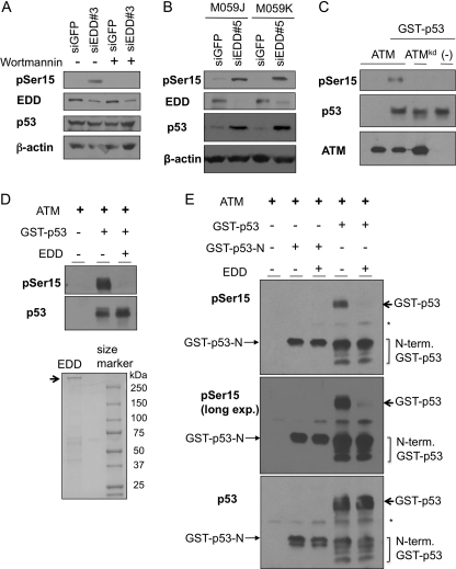
FIGURE 7.
EDD inhibits phosphorylation of p53 by ATM in vitro. A, WI38 cells infected with Ad-siGFP or Ad-siEDD#3 were left untreated or treated with wortmannin (20 μm). The cell lysates were harvest 1 h later and subjected to Western blot analysis. B, DNA-PK-deficient cell line M059J and its control M059K (DNA-PK wild type) were infected by Ad-siEDD#5 and Ad-siGFP. Three days later, cells were lysed, and p53 phosphorylation level at Ser15 was analyzed by immunoblotting. C, ATM and ATM kinase-dead mutant were immunoprecipitated from 293T cells, which were transfected with HA-ATM or HA-ATMkd construct. The ATM kinase assay was performed as described under “Experimental Procedures” and contained 200 nm GST-p53. Western blots were probed with an antibody against phospho-Ser15 of p53. D, 1 μg of EDD purified from 293T cells was incubated with 10 nm GST-p53 in 4 °C for 30 min first. Kinase assay was then performed as in C. E, 200 ng of EDD or 200 ng of BSA was incubated with 10 nm GST-p53 or GST-p53-N for 30 min. ATM kinase assay was then performed as in C. The top panel represents anti-pSer15-p53 immunoblot; the middle panel is a longer exposure (long exp.) of the same blot; and the low panel was probed with mixed p53 antibodies containing DO1, PAb1801, PAb240, and PAb421, which can recognize both GST-p53-N and GST-p53. * indicates nonspecific signals of IgG heavy chains. N-term GST-p53, N-terminal GST-p53.

