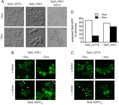
FIGURE 2.
Pik1 alters cell morphology but does not alter Ste5 localization. A, differential interference contrast image of TetO7-STT4, TetO7-PIK1, and TetO7-PIK1 kss1Δ cells treated with 10 μg/ml doxycycline for 15 h where indicated (+ Dox). B, GFP fluorescence of TetO7-PIK1 cells expressing pRS316-Ste5-(GFP)x3. Cells were treated with doxycycline for 15 h and 3 μm α factor pheromone for 90 min. Arrow heads indicate Ste5-GFP localized to shmoo tips. C, TetO7-STT4 cells treated as in B. Arrows indicate the absence of Ste5-GFP at shmoo tips. D, quantitation of the percentage of shmoos with polarized Ste5-GFP from B and C (n > 50 shmoos).

