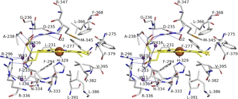
FIGURE 4.
Stereoview of heme binding site. The heme carbons are yellow sticks with iron atoms shown as brown spheres. Residues contributing to heme binding are shown as sticks with carbons in gray. The red stick represents the oxygen molecule and acts as the distal ligand. The water molecules are depicted as violet-purple spheres. The hydrogen bonds are depicted as dashed lines.

