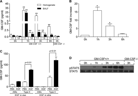
FIGURE 5.
KGF causes a rapid increase in GM-CSF protein and gene expression in alveolar type II cells and STAT phosphorylation in alveolar macrophages. Mice were given intranasal (i.n.) or intraperitoneal (i.p.) KGF or PBS. At the indicated time points, BAL was performed and lungs were harvested and homogenized. Panel A, GM-CSF levels in lung homogenate and BAL fluid (BALF) of KGF challenged GM-CSF+/+ and GM-CSF−/− mice were measured by ELISA. Data are mean ± S.E.; n = 3–4, **, p < 0.01; *, p < 0.05 for KGF versus 0 h unchallenged control. Panel B, GM-CSF gene expression was measured by real time PCR and compared with the constitutively expressed gene, β-actin. Data are mean ± S.E.; n = 3, *, p < 0.05, for KGF versus 0 h unchallenged control. Panel C, data represent GM-CSF levels in cell lysates of 1 million isolated alveolar macrophages (AMs) or type II cells (type II), harvested from mice 1 h after treatment with intranasal PBS or KGF in vivo. For in vitro experiments, 1 million alveolar macrophages or type II cells isolated from unchallenged mice and plated on plastic or Matrigel, respectively, were incubated with 200 ng/ml of KGF or PBS. After 24 h, the supernatants were harvested and GM-CSF levels were measured. Data are mean ± S.E.; n = 3. Panel D, strain-matched wild type (C57BL/6) and GM-CSF−/− mice were given with intranasal KGF or PBS. At the indicated time points, alveolar macrophages were isolated by BAL and STAT5 was immunoprecipitated from cell lysates using a rabbit polyclonal anti-STAT5 antibody. After SDS-PAGE and transfer to nitrocellulose, membranes were immunoblotted with anti-STAT5 and anti-phospho-STAT5 antibodies.

