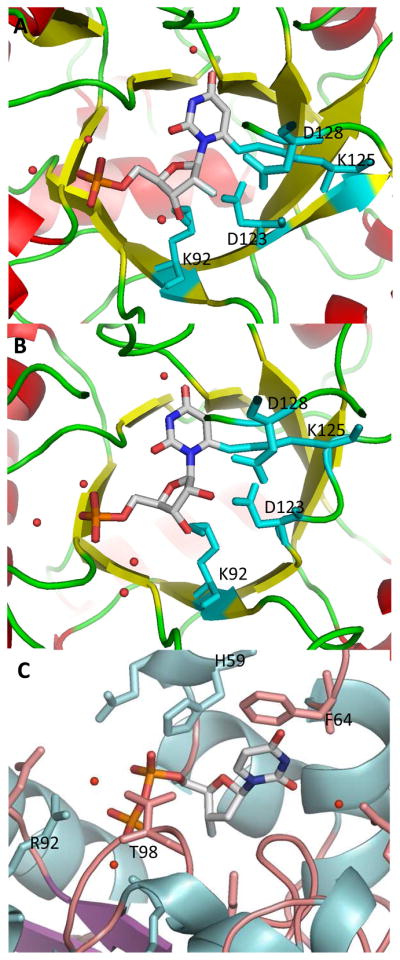
Figure 4.

(A) Co-crystal structure of human ODCase covalently modified by 2′-fluoro-6-iodo-UMP (33). (B) X-ray crystal structure of human ODCase covalently modified by 6-iodo-UMP (10) (pdb code: 3BGJ). (C) X-ray crystal structure of nucleoside diphosphate kinase bound by 3′-fluoro-UDP (pdb code: 1B99). Active site regions are shown with the ligand rendered according to atom type. The secondary structures of the proteins are shown, and only the important active site residues are shown in a capped stick representation. Water molecules are shown as red spheres.
