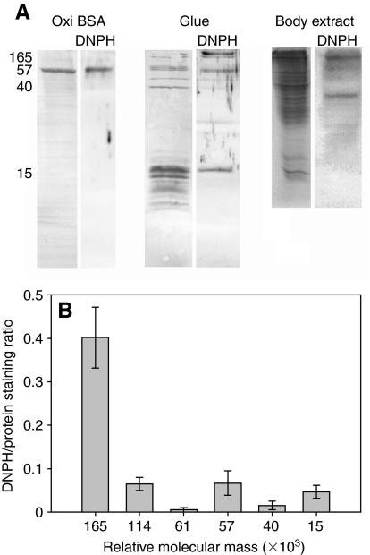
Fig. 1.
The extent of oxidation of the proteins in Arion subfuscus glue, based on anti-DNPH immuno-staining for carbonyl groups. (A) Sample of oxidized BSA (positive control), glue and whole body extract. Left lanes show each blot stained for total protein content using Coomassie Blue R-250. Right lanes (DNPH) show the corresponding anti-DNPH immune blots. The numbers on the left show relative molecular mass (×103). (B) Quantification of the extent of oxidation of selected proteins in the glue based on signal intensity. Values are the ratios of staining intensity of the immunostain relative to the staining intensity of Coomassie Blue for that protein on a duplicate blot (mean ± s.e.m., N=6). Oxidized BSA (positive control) had a DNPH/protein staining ratio of 0.37±0.04.

