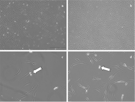
Fig. 1.
Mesenchymal cells in culture. The cells from a bone marrow or b fat tissue cultivated in α-modified Eagle's medium (αMEM) + 10% fetal calf serum (FCS) + antibiotic with change of medium every 3 or 4 days show similar aspects. Subculture at low density reveals small, round and refringent cells, which correspond to cells during division (white arrow) for c MSCs as well as for d ASCs. Magnification 40× (a and b) or 100× (c and d)

