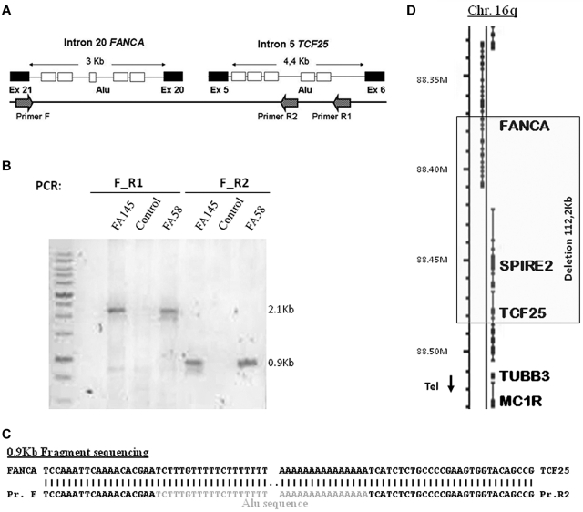
Figure 4.
PCR amplification of a fragment containing the breakpoint of ex1-20del. (A) Position of primers used, exons (black boxes), and Alu sequences (white boxes) found in regions flanking both breakpoints. (B) Fragments amplified by PCR in 2 patient carriers of the deletion (FA145 and FA58) and a control (not carrying this deletion) using 2 sets of primers. (C) Sequence of the 0.9-kb fragment containing the breakpoint (only relevant parts are shown). Sequence (bottom line) is aligned with FANCA and TCF25 (top line). (D) Scheme of the region affected by the deletion in chromosome 16q and genes included.

