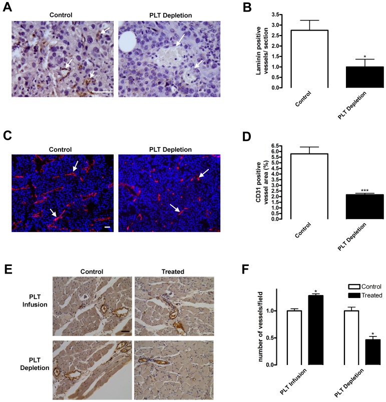
Figure 3.
Blood vessel density is diminished on platelet depletion. (A-D) Subcutaneous B16-F10 tumors were implanted in mice undergoing platelet (PLT) depletion or injected with control rat IgG. After 9 days of growth, tumors were excised and sectioned. (A-B) Tumor sections were stained for laminin to visualize blood vessels. Laminin-positive vessels were quantified and represented as mean vessel number per section ± SEM (n = 6). (C-D) Frozen tumor sections were stained for CD31+ vessels (red) and 4,6-diamidino-2-phenylindole (blue). CD31+ blood vessels were quantified and represented as mean percentage of area ± SEM (n = 24). Arrows indicate vessels within the tissues. (E-F) Platelets were depleted or infused in mice directly after hindlimb ischemia surgery. Muscles from mice injected with PBS (PLT Infusion, Control) or 3 × 10⁹ platelets (PLT Infusion, Treated) or rat IgG (PLT Depletion, Control) or rat antimouse GPIbα (PLT Depletion, Treated) were sectioned 14 days after surgery and immunostained for von Willebrand factor-positive blood vessels. Vessels were quantified and represented as mean vessel number per field ± SEM (n = 5). Scale bars represent 50 μm. *P < .05 vs control (Student t test). ***P < .005 vs control (Student t test).

