Figure 5.

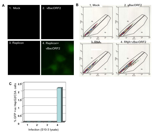
Infectivity assay with naïve HepG2 cells. (A) FM, showing expression of GFP in virion (S10-3 lysate)-infected cells (~5%), at day 6. (B) FACS plot, showing GFP expressing cells at day 6 post-infection. (C) %GFP positive cells in B, determined by FACS.
