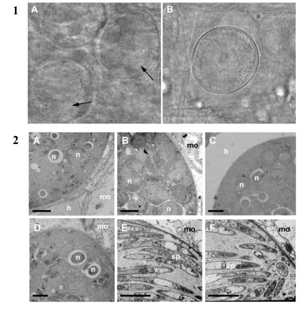
Figure 2.
Δpcrmp3 and Δpcrmp4 sporozoites develop normally but remain within mature oocysts. 1) At day 18 post infection, WT oocysts (A) contain few sporozoites and show areas of degradation (arrowed), whilst sporozoites within Δpcrmp3 oocysts (B) are radially-aligned and are retained within an intact cell wall. Scale = 100 mm. 2) WT (A), Δpcrmp3 (B) and Δpcrmp4 (C) oocysts are found in mosquito guts 10 days post infection, developing between the mosquito epithelium (mo) and haemolymph (h). Several nuclei (n) in developing sporozoites are visible. No morphological differences are observed. At day 18 post infection, WT oocysts contain few nuclei (D) whilst most Δpcrmp3 (E) and Δpcrmp4 (F) mature oocysts are full of sporozoites (sp). Scale = 5 μm.

