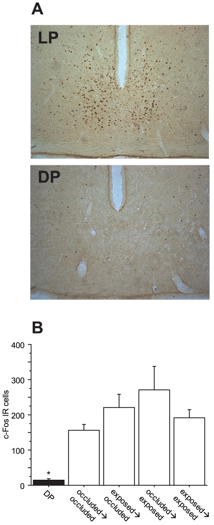
Figure 5. Light-induced c-fos expression in the SCN.
A Representative images of DAB-labeled Fos-IR cells in SCN coronal sections from 2 Syrian hamsters. B Mean (± s.e.m.) number of c-Fos immunoreactive (IR) cells in both SCN 90 minutes after the delivery of a dark pulse (DP) or a light pulse (LP) at CT19 (see methods for details) to an eye that had been either exposed or occluded from weeks 1–10 and from weeks 11–38. * p<0.0001 vs. all other groups.

