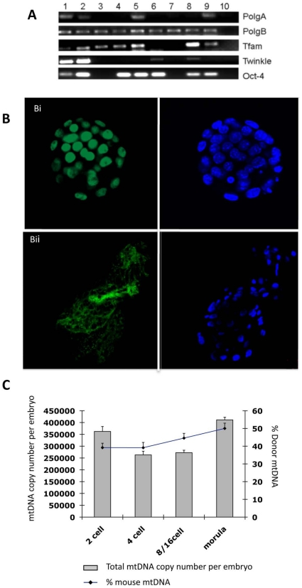
Figure 5. Analysis of pluripotent and mtDNA replication factors and mtDNA content in mtDNA-depleted, ESC supplemented-iSCNT embryos.
(A) RT-PCR analysis for gene expression of PolgA, PolgB, Tfam, Twinkle and Oct4 in single cloned embryos and ESCs. Lane 1–10 ESCs; Lane 2–20 ESCs; Lane 3–2-cell embryo; Lane 4–4-cell embryo; Lane 5–4-cell embryo; Lane 6–8-cell embryo; Lane 7–8-cell embryo; Lane 8-morula; Lane 9-blastocyst; Lane 10-Negative control. (B) Distribution of Oct-4 in an in vivo fertilised-derived blastocyst (i) and a murine-porcine cloned blastocyst (ii). Oct-4 and DAPI are shown in green and blue, respectively. (C) Total mtDNA copy number and percentage of donor cell mtDNA contribution in iSCNT embryos supplemented with murine ESC extract containing mitochondria.

