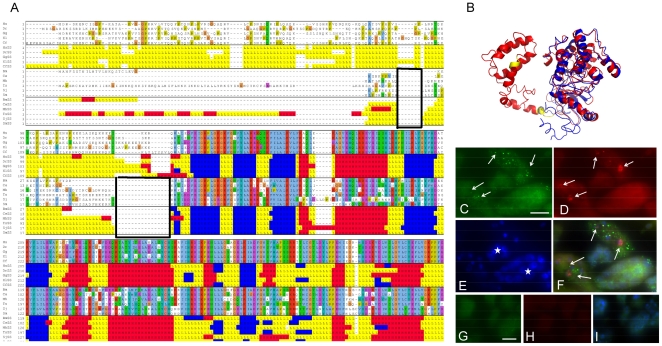
Figure 3. Characterization of O01427/Q19126.
A. Multiple sequence and secondary structure alignment of vertebrate reference sequences with selected nematode sequences. Within the secondary structure alignments, the random coiled regions are shown in yellow, the beta sheets are shown in blue and helices are shown in red. The two boxed regions show the deletions in the worms relative to the vertebrates. B. Predicted 3D structure of O01427 (H. sapiens protein in the orthologous group (red), B. malayi protein (blue), and indels (yellow)). C. Granular staining (arrows) for Q19126 [XP_00189449.1] mRNA in the cytoplasm of morula stage embryos in the midbody region of a female B. malayi. The biotin labeled probe was detected using AlexaFluor 488-labeled streptavidin (green). D. Identical section as in C showing granular staining (arrows) for O01427 [XP_001892118.1] mRNA in the same embryos. The digoxygenin labeled probe was detected using a Rhodamin conjugated anti-digoxygenin antibody (red). e. Identical section as in c but DAPI stain (blue) showing differential degrees of chromatin condensation in the embryos. f. Overlay of c-e showing co-localization of mRNA expression of Q19126 and O01427 (arrows) especially in embryos with less densely condensed chromatin. Hybridization of sense probe for Q19126 (g.) and O01427 (h.) on a serial section to c showing the absence of a specific labeling. i. Overlay of g and h including a DAPI stain (blue) showing the morula stage embryos with different degrees of chromatin condensation but the absence of a specific hybridization signal. Scale bar 10 µm.

