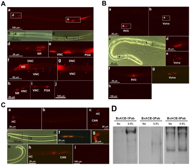
Figure 1. The localization pattern (A, B and C) and solubilization feature (D) of the three BxAChEs.
(A) BxACE-1. Nematodes from (a) to (e) were female adults, and those from (f) to (j) were larvae. a, BxACE-1 is shown around the pharynx (left white box), the putative ventral nerve cord (VNC, right white box) and the tail. b and c, The head and tail regions photographed using an optical microscope. The black boxes indicate the regions in which BxACE-1 was observed. d and e, The head and tail regions of the nematode. BxACE-1 was observed at the putative nerve ring (NR) located around the pharynx (d), VNC and the putative preanal ganglia (PGA) regions (e). f and g, The head and middle body regions of the nematode. BxACE-1 was observed at the putative NR, VNC and dorsal neuron cord (DNC). The red triangles indicate BxACE-1 in the VNC and DNC. h and i, Positive control. BxACE-1 was observed at the putative NR, VNC and PGA regions. (h) and (i) are the head and tail regions, respectively. j, Negative control, in which no signal was detected. (B) BxACE-2. The nematodes were female adults. a and b, BxACE-2 was observed around the post-NR (a) and putative vulva (b). c, Nematodes of (a) and (b) photographed using an optical microscope. The black boxes indicate the regions in which BxACE-2 was detected. d and e, Detailed photographs of white boxes in (a) and (b). f and g, Positive control. BxACE-2 was observed around the post-NR (f) and putative vulva (g). h, Negative control, in which no signal was detected. (C) BxACE-3. The nematodes were male adults (a–e and g–i) and larvae (f). a, BxACE-3 was observed at putative hypodermal cells (HC) located at the end of head. b, BxACE-3 was observed at the putative intestinal regions. c, BxACE-3 was observed at the putative canal-associated neuron (CAN), the DNC and HCs at the tip of the tail. d, Nematodes of (a) and (b) photographed using an optical microscope. The black boxes indicate the regions in which BxACE-3 was detected. e, Nematode of (c) photographed using an optical microscope. f, g and h, Positive controls. BxACE-3 was observed at the putative intestinal region (f), (h), HCs of tail (g), and HCs of head and intestinal regions (h). i, Negative control, in which no signal was detected. (D) Detection of each BxACE using corresponding anti-BxACE polyclonal antibody (BxACEPab) from the B. xylophilus crude protein samples extracted in the presence (lane 1) or absence (lane 2) of Triton X-100.

