Figure 1.
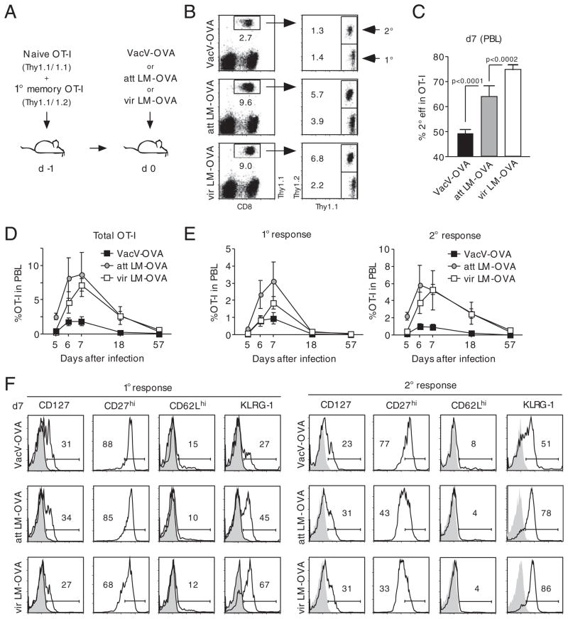
Pathogens differentially impact 2° effector CD8+ T-cell expansion and phenotype. (A) Experimental setup: 5 × 10² Thy1.1 naïve and 1 × 10⁴ Thy1.1/1.2 1° memory OT-I cells were mixed and injected into naïve Thy1.2 hosts (n = 8/group). Mice were infected with either 3 × 10⁶ PFU VacV-OVA i.p., 5 × 10⁶ CFU att LM-OVA or 5 × 10⁴ vir LM-OVA i.v. (B) Thy1.1+ OT-I cells were identified in PBL samples 7 days p.i. and 1° and 2° effector OT-I cells were distinguished by Thy1.1/1.2 costaining. Numbers show the percentage of the OT-I cell populations in the PBL population of representative mice. (C) Percentage (mean+SEM; n = 8 mice/group) of 2° effector CD8+ T cells in the total OT-I cell population in PBL 7 days p.i. (D) Kinetics of the combined 1° and 2° effector OT-I cell responses in PBL. (E) Kinetics of the 1° (left graph) and 2° (right graph) OT-I cell response in PBL. Numbers show mean±SEM (n = 8). (F) PBLs of all mice (n = 8/group) 7 days p.i. were pooled and the phenotype of 1° (left panel) and 2° (right panel) effector OT-I cells was analyzed. Numbers show the percentage of marker-positive OT-I cells, and shaded histograms represent isotype controls. Data are representative of two independent experiments.

