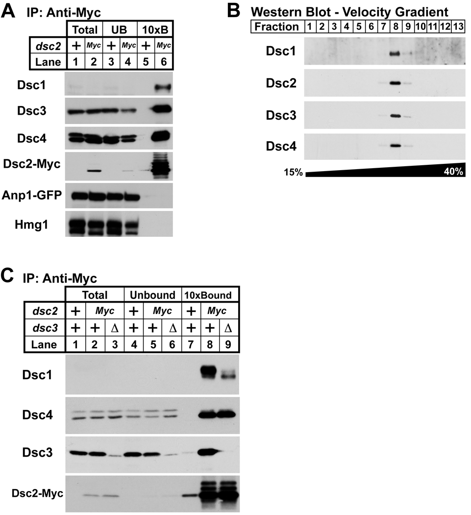
Figure 3. Dsc proteins form a complex.
(A) Digitonin solubilized extracts from anp1-GFP and dsc2-Myc anp1-GFP cells were prepared, and proteins associated with Dsc2-Myc were immunopurified with anti-myc IgG-9E10 monoclonal antibody. Equal amounts of total (lanes 1 and 2) and unbound fractions (lanes 3 and 4) along with 10× bound fractions (lanes 5 and 6) were immunoblotted using anti-Dsc1 IgG, anti-Dsc3 serum, anti-Dsc4 serum, rabbit anti-Myc IgG, anti-GFP and anti-Hmg1 IgG. (B) Eluate from tandem affinity purification using dsc1-TAP cells was subjected to velocity centrifugation on a 15%–40% sucrose gradient. Fractions were analyzed by immunoblot using anti-Dsc1 IgG, anti-Dsc2 serum, anti-Dsc3 serum, and anti-Dsc4 serum. (C) Digitonin solubilized extracts from anp1-GFP, dsc2-Myc anp1-GFP, and anp1-GFP dsc2-Myc dsc3Δ cells were prepared, and proteins associated with Dsc2-Myc were immunopurified using anti-myc IgG-9E10 monoclonal antibody. Equal amounts of total (lanes 1, 2 and 3) and unbound fractions (lanes 4, 5 and 6) along with 10× bound fractions (lanes 7, 8 and 9) were immunoblotted using anti-Dsc1 IgG, anti-Dsc4 serum, anti-Dsc3 serum, and rabbit anti-Myc IgG. For (A) and (C), + denotes wild-type allele.

