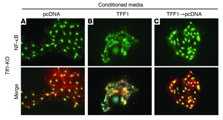
Figure 7. TFF1 negatively regulates NF-κB activity in primary epithelial cells.
Ex vivo immunofluorescence assay was performed on primary gastric epithelial cells isolated from the antrum of Tff1-knockout mouse. (A) Nuclear immunostaining of NF-κB–p65 (green fluorescence) was detected in cells that were treated with conditioned media from AGS-pcDNA cell line for 48 hours. (B) Primary gastric epithelial cells treated with conditioned media from AGS-TFF1 cell line for 48 hours displayed loss of nuclear immunostaining of NF-κB–p65. (C) On the other hand, gastric epithelial cells treated with conditioned medium from the AGF-TFF1 cell line for 24 hours, followed by replacement of this medium with conditioned medium from AGS-pcDNA cell line for another 24 hours restored the nuclear localization of NF-κB–p65. ZO1 (red) immunostaining was used as an epithelial cell marker, and DAPI (blue) was used as a nuclear counterstain. The results represent 1 of 3 independent experiments. Original magnification, ×40.

