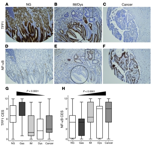
Figure 9. Immunohistochemistry for TFF1 and NF-κB in human samples.
(A–F) Immunohistochemical staining for TFF1 and NF-κB in serial tissue sections from human gastric mucosa with normal histology (NG; A and D), intestinal metaplasia and dysplasia (IM/Dys; B and E), and adenocarcinoma (C and F). A progressive decrease of TFF1 expression was observed from normal mucosa to adenocarcinoma, along with a progressive increase in p–NF-κB–p65 expression. The circular and rectangular areas demonstrate an inverse relationship in the immunostaining of TFF1 and p–NF-κB–p65 in serial sections from the same tissue. Original magnification, ×20. (G and H) The graphs summarize the immunohistochemical staining results on gastric tissue microarrays.

