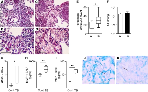
Figure 5. MMP-1 drives matrix destruction in TB granulomas of transgenic mice.
Mice expressing human MMP-1 in activated macrophages and wild-type mice littermates were infected with M. tuberculosis H37Rv. At 130 days, mice were sacrificed. (A and B) In wild-type mice, alveolar walls are intact on Masson’s Trichrome staining. (C and D) In contrast, in MMP-1–expressing mice, alveolar walls have been destroyed within areas of pneumonia. (E) Alveolar wall integrity in regions of pneumonia was scored by a pathologist blinded to the mouse genotype. Increased alveolar wall destruction was demonstrated in mice expressing MMP-1. (F) No significant difference in colony counts was demonstrated at 230 days after infection. (G) Relative MMP1 mRNA levels are increased in infected transgenic mice. (H) MMP-1 protein concentration is increased in BALF and (I) lung homogenates of M. tuberculosis–infected mice compared with those of uninfected transgenic mice. (J and K) Acid-fast bacilli are demonstrated in infected macrophages in (J) wild-type and (K) MMP-1–expressing mice. High resolution images are shown in Supplemental Figure 5. For each experiment, a minimum of 4 mice per group were studied, and a total of 4 separate infection experiments were performed. For box-and-whisker plots, the box outline represents the 25th and 75th percentiles, the central line represents the median value, and the whiskers represent minimum and maximum values. *P < 0.05, **P < 0.01 by Student’s t test. Scale bars: 50 μm for all panels.

