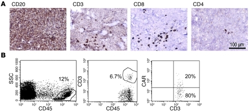
Figure 3. Detection of CAR+ T cells in a skin tumor biopsy.
(A) Immunohistochemical examination (diaminobenzidine with hematoxylin counterstaining) of a punch biopsy of a lymphoma skin lesion from patient number 5 at 2 weeks after T cell infusion showed that tumor cells were CD20+ (shown), CD10+, BCL2+, and BCL6+, consistent with involvement by follicular lymphoma with large cell transformation. Scattered CD3+ CD8+ cells infiltrated the tumor. Of note, the infused CAR.CD19-28ζ+ product consisted of 85% CD8+ cells. Scale bar: 100 μm. (B) FACS analysis of a cell suspension obtained from a fragment of the tumor biopsy. Viable cells represented approximately 45% of the preparation. The far left panel shows the gate on CD45+ cells, which represented 12% of the viable cells. The middle panel shows the CD3+ lymphocytes infiltrating the tumor, which accounted for 6.7% of CD45+ cells (0.8% of all viable cells). The far right panel illustrates that 20% of the gated CD3+ lymphocytes cells coexpressed the CAR, as assessed by the Fc-Cy5 monoclonal antibody, which binds to the IgG1-CH2CH3 spacer region of the CD19-specific CARs (~0.16% of all viable cells). SSC, side scatter.

