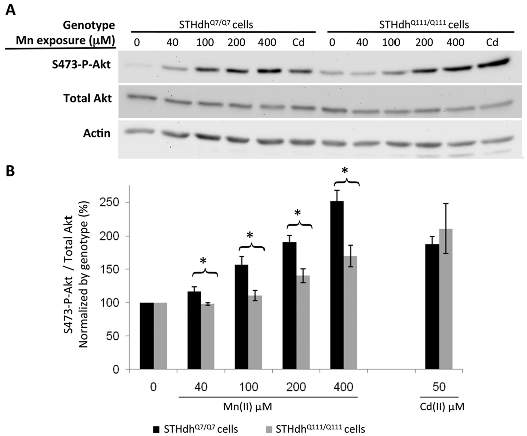
Fig. 5.
Diminished manganese-dependent Akt phosphorylation in HD striatal cells. (A) Lysates harvested from STHdhQ7/Q7 (wild-type) or STHdhQ111/Q111 (mutant) cells after 3 hours of manganese chloride exposure were analyzed by western blot for phosphorylated Akt (S473-P-Akt), total Akt and actin. Representative blots are shown. (B) Quantification of S473-P-Akt/total Akt expression in striatal cell lines. Mean values were normalized by genotype to the vehicle-only control (± SEM, n=4 independent samples). Significant differences in protein levels (* p<0.05 post-hoc t-test) between genotypes are indicated for each exposure.

