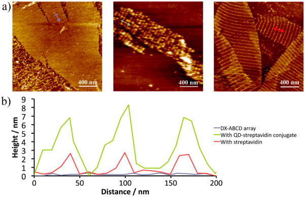
Figure 2.
a) From left to right: AFM images of DX-ABCD array alone with each A tile bearing a biotin; the biotinylated DX-ABCD array incubated with STV-QD conjugate; and same array incubated with streptavidin only; b) Cross section analysis of the AFM images. Each trace corresponds to the same colored arrow in a).

