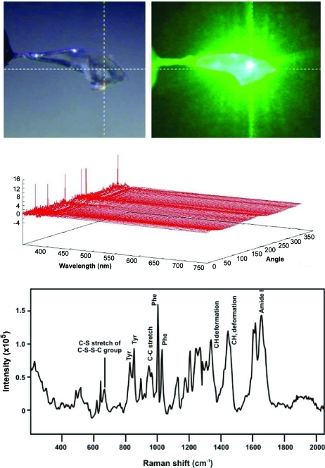
Figure 7.
An example of single-crystal electronic absorption and Raman spectroscopy collected from the same Zn2+ insulin crystal (Stoner-Ma et al., 2011 ▶). The top two panels show the crystal illuminated with visible light (left) and with the 532 nm laser (right). The middle panel shows the 72 electronic absorption spectra as a function of crystal rotation angle. The bottom panel shows the Raman spectra collected using 6 mW of 532 nm laser excitation and 50 s total acquisition time. This particular crystal morphology has less dependence on finding the ‘best’ or flattest face.

