Figure 5.
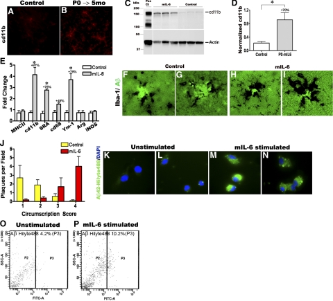
mIL6-induced persistent microglial up-regulation results in efficacious plaque clearance. A, B) Analysis of cd11b/Mac in mIL-6-expressing P0 → 5 mo TgCRND8 mice (n=3–4/group). Representative image showing up-regulation of cd11b/Mac in mIL-6-expressing TgCRND8 mice (B) compared with controls (A) detected by immunofluorescent staining on free-floating fixed sections. View ×200. C) Representative immunoblot analysis of cd11b in RIPA soluble brain extracts from mIL-6-injected P0 → 5 mo TgCRND8 mice compared with age-matched controls. D) Quantitative analysis of cd11b immunoblotting after normalizing to β-actin levels. *P < 0.05. E) Quantitative RT-PCR analysis of levels of microglial markers in mIL-6-expressing mouse forebrain. Data (fold change over control) represent average values obtained by QPCR on 3-mo-old mice injected with AAV1-EGFP (control) or AAV1-mIL-6 on P0; 2 independent experiments; n = 4 mice/group. *P < 0.001; 2-way ANOVA with Bonferroni’s posttests. F–I) Representative images of thioflavin-S-stained Aβ plaques (fluorescent green labeling) decorated with Iba-1 immunoreactive microglia (black immunostain) in mIL-6-expressing P0 → 5 mo TgCRND8 mice (H, I) and controls (F, G). View ×400. J) Quantitative analysis of the extent of Iba-1 immunodeposits circumscribing individual plaques shows increased association of activated microglia with plaques in mIL-6-expressing P0 → 5 mo TgCRND8 mice compared with controls (n = 4–5 mice/group). K–N) mIL-6-treated primary microglia appear to be more efficient in the uptake of fAβ42-Hilyte488 (green fluorescence, M, N) compared with unstimulated glia (K, L). Blue fluorescence indicates DAPI-stained glial nuclei. Data from 2 independent experiments; view ×600. O–P) Microglial cells with internalized Aβ42-Hilyte 488 (FITC channel on x-axis) were quantified by FACS. Percentage of positive cells in mIL-6 stimulated microglial cells was 10.2% (P, P3) compared with 4.2% in control unstimulated cells (O, P3).

