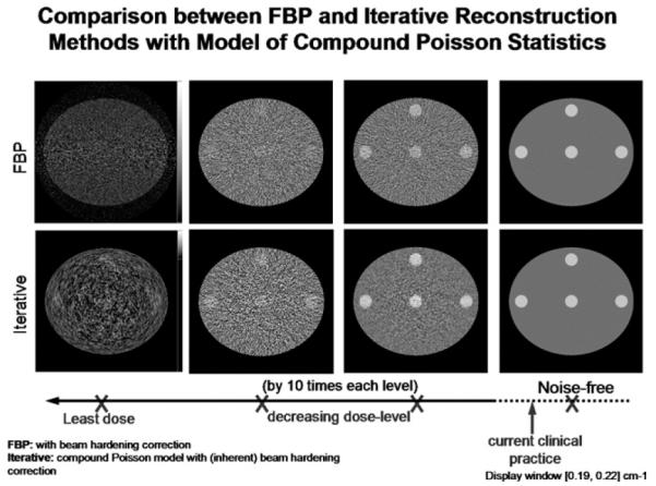
Fig 1.

A simulation study comparing reconstructed images at different radiation dose levels. (Top row) Conventional FBP reconstruction; (bottom row) statistical IR [4]. The images in the fourth column are from noise-free projection data. The dose increases by 10 times each from the first to the third column. The advantage of statistical IR methods over FBP methods is demonstrated at lower radiation dose levels (images in the 1st-3rd column). The clinical dose level is shown schematically on the horizontal dose axis.
