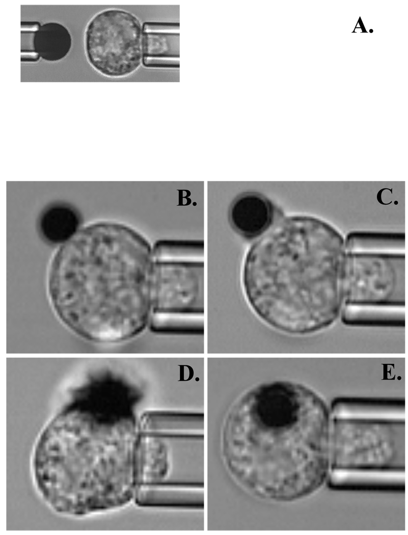
Figure 1.
Series of video microphotographs showing the experimental setup for the micropipette experiment. A. Initial setup. Left pipette is holding ICAM-1 coated bead (4.5µm in diameter) and right pipette is holding human neutrophil. During the experiment the adhesion probability between the neutrophil and ICAM-1 coated bead was measured. B–E. Time dependent IL-8 bead (2.8 µm in diameter) engulfment. Snapshots are taken at: B. 0 seconds (initial contact), C. 20 seconds, D. 70 seconds, E. 320 seconds – after IL-8 bead attachment to the neutrophil.

